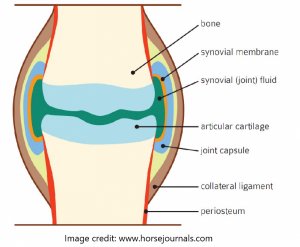
This above image simply shows an equine joint. By itself, it’s nothing spectacular, but sadly enough the equine industry often deals with a joint as if it only exists of bone. I often hear “the horse is lame, but the X-rays are clean so the problem can’t be there.” X-rays are an important part of the tools we use to assess joint health, but as this image shows there is more to a joint then what’s visible on an X-ray. Practically, an X-ray will show a detailed image of the bone in a joint, bone lining and bony pieces in the joint space (chips). What it won’t show is any of the soft tissue (joint capsule, ligaments, or synovia) and it won’t even show the cartilage until it’s too late and the cartilage is gone and replaced with bony spurs and changes which we then call arthritis.
Changes in balance, soundness, lack of push, less elevation, mild swelling (especially after work), heat, tenderness at touch and foot placement in rest are all factors that can indicate there is a joint problem. It’s great and important that the X-rays are “clean” but that doesn’t mean there is no problem in or near that joint!